Before You Start
Standard Setup
CVS Examination Checklist
- Introduce yourself, explain exam, obtain consent
- Ensure privacy, warmth, good lighting. Wash hands.
- Stand on the right side of the patient
- Position patient semi-recumbent at 45°
- Expose anterior chest to the umbilicus (above waist)
- General observation: conscious, alert, oriented — looks well or ill — breathless, cyanosed, distressed
- Record vitals: RR, Temp, BMI, BP, Pulses
History Taking
Chief Complaints
A — Chest Pain

| Angina | MI | Aortic Dissection | Acute Pericarditis | |
|---|---|---|---|---|
| Site | Retrosternal | Retrosternal | Retrosternal, Intrascapular | Retrosternal or left-sided |
| Onset | Gradual 1–2 min | Rapid few mins | Very sudden | Gradual; postural change aggravates |
| Character | Constricting, heavy | Constricting, heavy | Tearing, ripping | Sharp, stabbing, pleuritic |
| Radiation | Arms, neck, epigastrium | Arms, neck, jaw | Back, between shoulders | Lt shoulder or back |
| Relieving | Rest, nitrates | NOT relieved by rest | Nothing relieves | Sitting up, leaning forward, NSAIDs |
| Associated | Breathlessness | Sweating, nausea, Angor Animi | Syncope, focal neuro signs | Flu-like prodrome, fever |
B — Shortness of Breath (Dyspnea)


Breathlessness: Onset → Diagnosis
- Minutes: PE, Pneumothorax
- Hours–days: Pneumonia, Asthma
- Weeks–months: Anaemia, Pleural effusion
- Months–years: COPD, Pulmonary fibrosis
MRC Breathlessness Scale
- Grade 1: Hurrying on level/hill
- Grade 2: Walking with peers on level
- Grade 3: Walks slower than peers
- Grade 4: Stops after 100m
- Grade 5: Too breathless to leave house
- Grade 5b: Too breathless to wash/dress
C — Palpitations

| Extrasystoles | Sinus Tachy | SVT | AF | VT | |
|---|---|---|---|---|---|
| Onset | Sudden | Gradual | Sudden, with 'jump' | Sudden | Sudden |
| Character | 'Jump', missed beat | Regular, fast, pounding | Regular, fast | Irregular, fast | Regular, fast |
| Associated | Nil | Anxiety | Polyuria, chest tightness | Breathlessness | Presyncope, syncope |
| Severity | Mild | Mild–moderate | Moderate–severe | Variable | Often severe |
D — Syncope

| Postural Hypotension | Vasovagal | Arrhythmias | Mechanical | |
|---|---|---|---|---|
| Onset | On standing | Sudden | Sudden | — |
| Prodrome | Lightheadedness, tinnitus, dark vision, sweating | Same as left | Palpitations, chest pain, SOB | — |
| Duration | 1–2 mins | <60 seconds | — | — |
| DDx | Hypovolemia, drugs (diuretics) | Vasovagal attack | VT, complete heart block | Severe AS, HOCM, PE |
E — Lower Limb Swelling


Hands
Peripheral Signs
Hands examination — nails, palms, dorsum


Inspection — What to Look For
- Nails: Tobacco stain · peripheral cyanosis · clubbing · splinter haemorrhages
- Palms: Janeway spots · Osler nodes · pallor · palmar erythema
- Dorsum: Petechial rash · tendon xanthomata
- IV drug use sites
- Tremor (fine + flapping)
- Temperature + sweaty or dry


Palpation
- Temperature (wet / dry)
- Capillary refill — normally <2 seconds
- Pulses (see next section)


Face
Facial Signs

Face examination — xanthelasmata, malar flush, central cyanosis, corneal arcus
Eyes
- Xanthelasmata on eyelids
- Conjunctival pallor and petechial haemorrhage
- Corneal arcus on iris
- Fundoscopy — DM/HTN changes, Roth spots
Cheeks & Mouth
- Malar flush — suggests mitral stenosis
- Central cyanosis under the tongue
- Peripheral cyanosis on the lips
- Dental caries (infective endocarditis risk)
Pulse Examination
Rate · Rhythm · Volume · Character

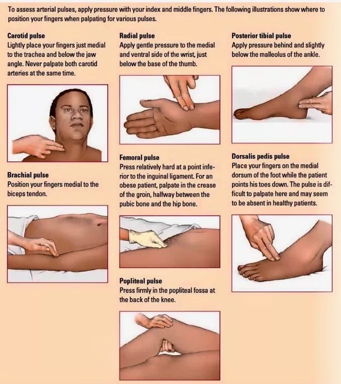
Radial pulse — feel just lateral to flexor carpi radialis tendon with pads of 3 fingers
Comment on: Rate · Rhythm · Volume · Character · Compressibility
— Then radio-radial delay · radio-femoral delay · collapsing pulse · pulse deficit
Pulse Locations


⚠️ Carotid Pulse — Critical Safety Rule
NEVER feel both sides simultaneously — risk of cerebral ischaemia.
Auscultate for bruit on BOTH sides while patient holds breath.
| Pulse | Location | Comment On |
|---|---|---|
| Radial | Just lateral to flexor carpi radialis tendon | Rate, rhythm, volume, character, radio-radial delay, collapsing pulse, deficit |
| Brachial | Medial to biceps tendon in antecubital fossa | Volume, character, compressibility |
| Carotid | Between larynx and anterior border of SCM | ONE SIDE ONLY — auscultate for bruit |
| Femoral | Midpoint of inguinal ligament (ASIS → pubic symphysis) | Bruits |
| Popliteal | Press firmly behind 30° flexed knee, both hands | Palpable / not palpable |
| Post. Tibial | 2cm below and behind medial malleolus | Palpable / not palpable |
| Dorsalis Pedis | Lateral to extensor hallucis longus, against navicular | Absent in 2–3% of healthy individuals |
Pulse Rate & Rhythm


Pulse Character
Slow Rising
Gradual upstroke with reduced peak occurring late in systole
Severe aortic stenosis
Collapsing (Water Hammer)
Peak arrives early → rapid fall; wide pulse pressure (systolic – diastolic >80 mmHg)
Severe aortic regurgitation, PDA
Pulsus Bisferiens
Double systolic peak separated by a mid-systolic dip
AS + AR combined, HOCM
Pulsus Alternans
Beat-to-beat variation in volume with normal rhythm
Advanced heart failure
Pulsus Paradoxus
Exaggerated normal variation with breathing (>10 mmHg fall in systolic on inspiration)
Cardiac tamponade, pericardial constriction, acute severe asthma
Blood Pressure
Korotkoff · Classification
BP Measurement Technique
- 1Rest 5 minutes, no tight clothing
- 2Support arm at heart level
- 3Apply proper-sized cuff — bladder over brachial artery
- 4Palpate brachial artery → inflate to 30 mmHg above impalpable pulse
- 5Deflate slowly — Phase 1 = systolic, Phase 5 = diastolic
- 6Measure both arms; record site and position
| BP Category | Systolic (mmHg) | Diastolic (mmHg) |
|---|---|---|
| Optimal | <120 | <80 |
| Normal | <130 | <85 |
| High Normal | 130–139 | 85–89 |
| HTN Grade 1 (mild) | 140–159 | 90–99 |
| HTN Grade 2 (moderate) | 160–179 | 100–109 |
| HTN Grade 3 (severe) | >180 | >110 |
Jugular Venous Pressure (JVP)
Right Atrial Pressure


Clinical clues to secondary hypertension

JVP Examination Sequence
- 1Position patient supine at 45°, pillow behind head, head slightly turned left
- 2Inspect: use tangential light → look for rapid inward movement, two waves per pulse
- 3Palpate: JVP is impalpable — compresses with neck pressure and disappears
- 4Special manoeuvres: respiration (↓ inspiration), lie flat (↑), sit up (↓), abdominojugular reflux
- 5Measure height: vertical distance from tip of visible pulsation to sternal angle. Normal <9 cmH₂O
JVP Waveforms
a
Atrial systole
Right atrial contraction. Absent in AF. Giant in tricuspid stenosis.
c
Tricuspid closure
RV contraction pushing tricuspid valve back into RA
x
x descent
Downward displacement of tricuspid ring during systole
v
Venous filling
Peak RA pressure before tricuspid opens. Giant in TR.
y
y descent
Commencement of ventricular filling. Prominent in pericardial constriction.
| Condition | JVP Finding |
|---|---|
| Heart failure | Elevation + sustained abdominojugular reflux >10 seconds |
| Pulmonary embolism / Tamponade | Elevation |
| Pericardial effusion | Elevation + flattened 'y' descent |
| Pericardial constriction | Elevation + Kussmaul's sign + prominent 'y' descent |
| SVC obstruction | Elevation + loss of pulsation (non-pulsatile) |
| Atrial fibrillation | Absent 'a' waves |
| Tricuspid stenosis | Giant 'a' waves (large, prominent) |
| Tricuspid regurgitation | Giant 'v' or 'cv' waves |
| Complete heart block | 'Cannon' waves |
Kussmaul's Sign
Paradoxical rise of JVP with inspiration
DDx: Pericardial constriction · Severe RV failure · Restrictive cardiomyopathy
Precordial Examination
Inspect · Palpate
Precordium examination — Inspection · Palpation · Auscultation
Inspection
What to Inspect
- 1From foot of bed: chest deformities (pectus excavatum, barrel chest) and body hair distribution
- 2From right side (lean forward to see left axilla): scars, masses, visible pulsations, dilated veins
- 3Visible pulsations — apex beat, parasternal pulsations
- 4Surgical scars — sternotomy, pacemaker insertion, chest drain sites
Palpation — Apex Beat
Apex Beat — Technique
- 1Find most lateral and inferior palpable pulse using entire palm
- 2Confirm location with 2 fingers — should be left 5th ICS, mid-clavicular line
- 3If impalpable → tilt patient to left lateral decubitus position
- 4If still impalpable → consider hyperinflated lungs (COPD) or dextrocardia
Impalpable
Hyperinflated lungs (COPD) or dextrocardia
Diffuse + displaced inferolaterally
LV dilatation
Forceful + undisplaced
LV hypertrophy
Double impulse
Hypertrophic cardiomyopathy
Tapping
Mitral stenosis (palpable S1)
Heave
Use the heel of your hand at the left parasternal area at end of expiration.
A heave suggests RV hypertrophy or dilatation / pulmonary HTN.
Auscultation
Heart Sounds · Murmurs


Auscultation Sequence
- 1Diaphragm — all 4 areas (Aortic, Pulmonary, Tricuspid, Mitral). Keep other hand on carotid. Ask to breathe deep at pulmonic for splitting. Auscultate left axilla (MR) and carotids while breath-held (AS).
- 2Sit and lean forward, hold breath → auscultate aortic area + Erb's point for early diastolic murmur of AR
- 3Bell over all 4 areas — S3, S4. Roll patient to left lateral position + bell for mid-diastolic murmur of MS

Heart Sounds
Normal Sounds
- S1 (Lub): Closure of mitral + tricuspid valves. Loudest at apex.
- S2 (Dub): Closure of aortic + pulmonary valves. Loudest at base.
- Physiological splitting of S2 on inspiration (normal)
- Fixed splitting → ASD
- Paradoxical splitting → AS / LBBB
Added Sounds
- S3: Normal in young; gallop in adults = heart failure
- S4: Always pathological → stiff ventricle (LVH)
- Opening Snap: After S2 → mitral stenosis
- Ejection Click: After S1 → valve stenosis
- Friction Rub: Pericarditis
Murmurs
Aortic Stenosis (AS)
Midsystolic ejection murmur
Best heard: 2nd right ICS (aortic area)
Radiates to: carotids
Harsh, crescendo-decrescendo
Radiates to: carotids
Harsh, crescendo-decrescendo
Mitral Regurgitation (MR)
Pansystolic murmur
Best heard: Apex
Radiates to: left axilla
Blowing, uniform intensity
Radiates to: left axilla
Blowing, uniform intensity
Aortic Regurgitation (AR)
Early diastolic murmur
Best heard: Erb's point (3rd–4th left ICS)
Patient: sitting + leaning forward + breath-held at end expiration
Soft, blowing, decrescendo
Patient: sitting + leaning forward + breath-held at end expiration
Soft, blowing, decrescendo
Mitral Stenosis (MS)
Mid-diastolic murmur
Best heard: Apex with BELL
Patient: left lateral position
Only murmur heard with the BELL
Patient: left lateral position
Only murmur heard with the BELL
"Normal S1 and S2 with physiological splitting of S2 on inspiration. No added sounds (S3, S4, opening snap, ejection click, friction rub). No murmurs."
— Normal Auscultation OSCE Comment · If murmur: location, radiation, timing, character, pitch
Lower Limb & Peripheral Vascular
PVD · Ulcers · Neurological
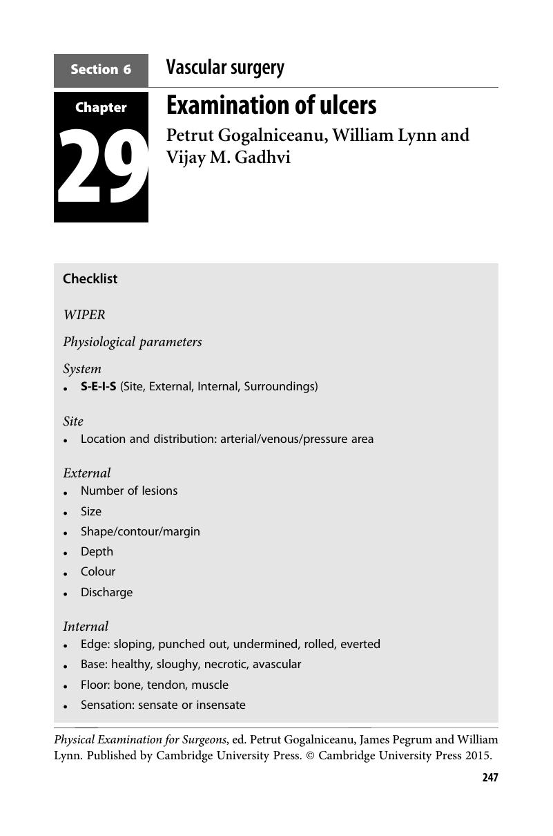
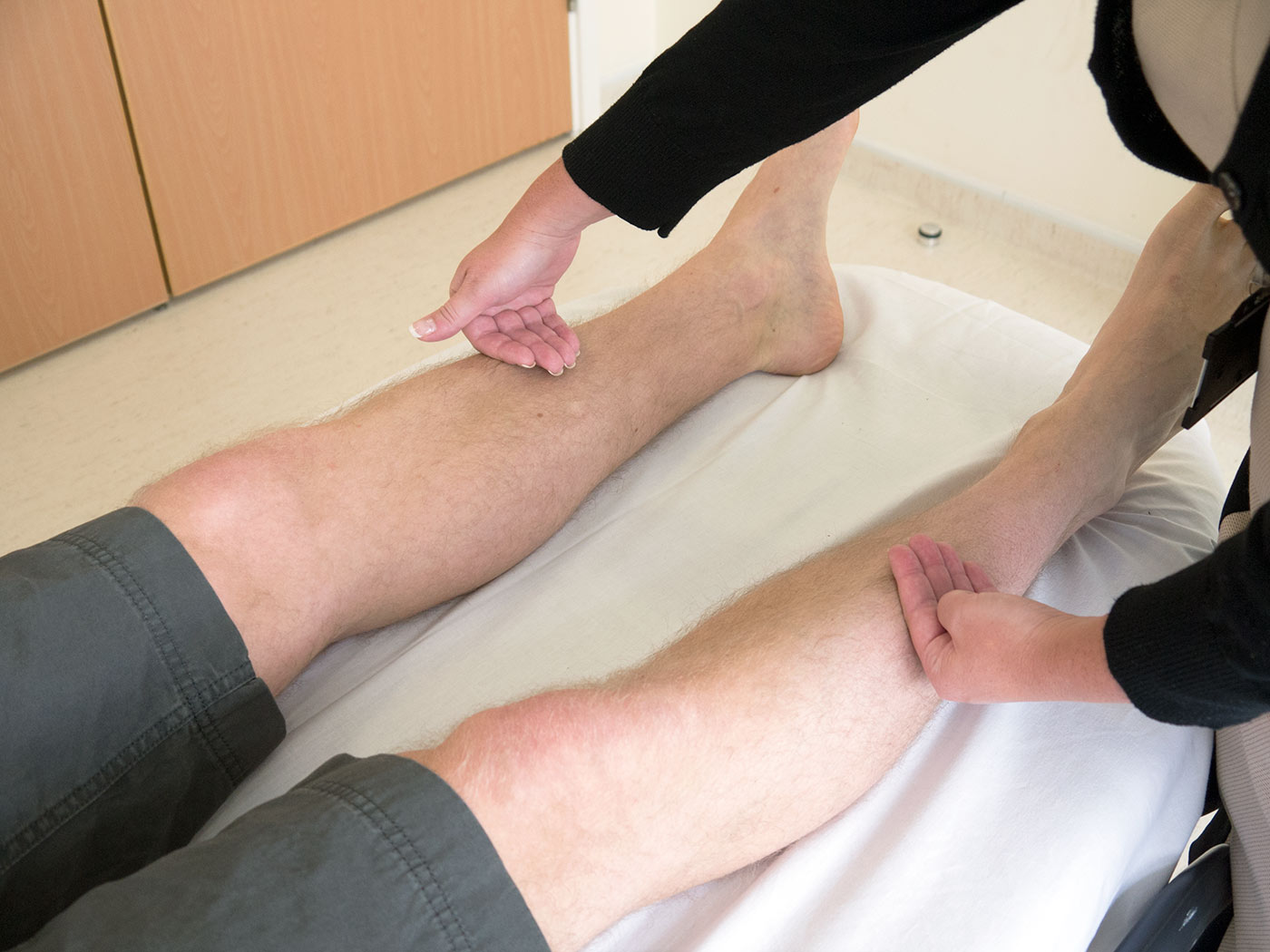
Inspection
Signs of Ischemia
- Amputated limb/toe · Thick hypertrophied nails
- Peripheral cyanosis · Pallor · Hair loss
- Dry/shiny skin · Decreased muscle bulk
- Black discoloration of toes (gangrene)
- Charcot's joint deformity
- Check heel and between toes for ulcers
Signs of Venous Insufficiency
- Visible distended / tortuous veins
- Redness or hyperpigmentation (haemosiderin)
- Thick skin · Obvious scars · Ulcers
- Obvious swelling
Ulcer Types & Sites
Arterial ulcer
Tip of toes, dorsum of foot — pale, deep, painful, punched-out
Tip of toes, dorsum of foot — pale, deep, painful, punched-out
Venous ulcer (long saphenous)
Medial side of leg (gaiter area)
Medial side of leg (gaiter area)
Venous ulcer (short saphenous)
Lateral side of leg
Lateral side of leg
Pressure ulcer
Over sole at pressure points
Over sole at pressure points
Palpation
Special Tests


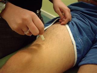
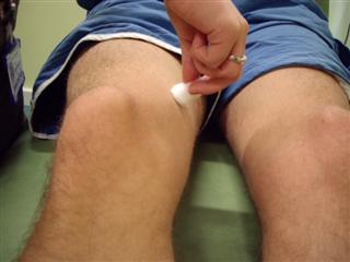
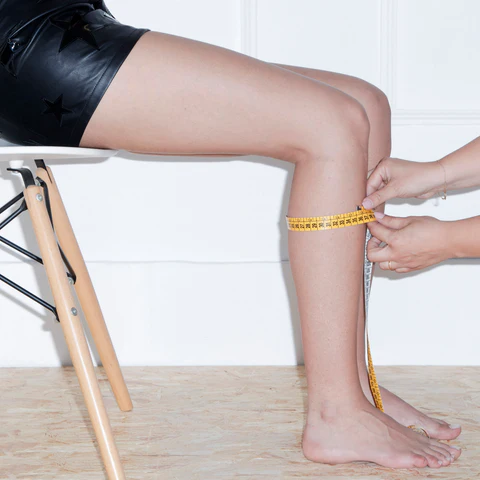
Buerger's Test (PAD)
- 1Raise legs to 45°, support for 2–3 minutes
- 2Watch for pallor + 'guttering' of superficial veins
- 3Sit patient up, hang legs over bed edge
- 4Cyanosis before returning pink → positive = PVD
Trendelenburg Test (Varicose Veins)
- 1Elevate leg — 'milk' veins toward groin
- 2Press sapheno-femoral junction (2–3 cm below + lateral to pubic tubercle)
- 3Ask patient to stand while maintaining pressure
- 4Veins fill on releasing → sapheno-femoral incompetence
Ankle-Brachial Index (ABI)
ABI Interpretation
>1.4
Calcification / vessel hardening
→ Vascular specialist
1.0–1.4
Normal
None
0.9–1.0
Acceptable
None
0.8–0.9
Some arterial disease
Treat risk factors
0.5–0.8
Moderate arterial disease
→ Vascular specialist
<0.5
Severe arterial disease
→ Vascular specialist (urgent)
Neurological Examination
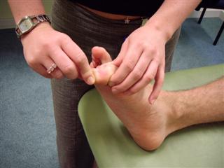
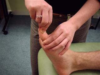
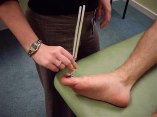
Neurological examination — light touch · pin-prick · vibration · proprioception · ankle jerk
To Complete the Examination
Always state these at the end of your OSCE presentation
• Lung bases — auscultate for crackles
• Hepatomegaly and ascites
• Lower limb and sacral oedema
• Examine inguinal lymph nodes and gait